
Report on the 2nd iCeMS-CiRA Classroom: Hands-On with Stem Cells!
August 16, 2010
On August 4 and 5, 2010, the Institute for Integrated Cell-Material Sciences (iCeMS) and the Center for iPS Cell Research and Application (CiRA) held the second iCeMS-CiRA Classroom "Hands on with Stem Cells", a laboratory exercise-based educational program for high school students. Additional sponsorship was provided by the Kyoto Prefectural Board of Education, as part of a higher-education outreach effort.
32 participants (out of 86 applicants from around the country) visited the iCeMS in spite of Kyoto's notorious midsummer heat.
One of the biggest goals of the program was to help the high school students acquire the tools of scientific inquiry. Workshop staff assisted them in formulating hypotheses, testing, and then attempting to explain and organize the results of experiments using embryonic and induced pluripotent stem (ES and iPS) cells.
The 32 students were divided into eight groups, each like an individual research team. Eight high school teachers, who had already participated in a similar program, acted as teaching assistants.
Day One (August 4, 2010)
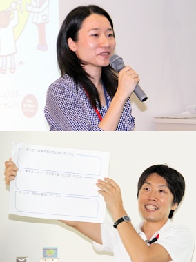
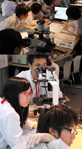
iCeMS Drs. Mizumachi (above) and
Kano briefing the students
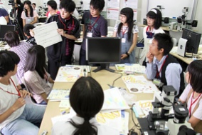
Students present discussion points
Mr. Takeuchi (above) and Ms. Ogura explaining
the science behind the experiments
Entering the world of stem cells
Using the Classroom Study Book as a starting point, students gained basic knowledge of stem cells and acquired the techniques to conduct experiments on them.
As it is important for students to learn how the techniques of discussion with teammates are an essential part of research, educational tools were prepared to promote debate and help the students discuss topics such as "Making a Hypothesis", "Cells", "The Genome", "Stem cells", and "Bioimaging".
Hands-on experiences were also provided, such as the molding ES cells out of clay, using micropipettes, and using phase invert microscopes.
Day Two (August 5, 2010)
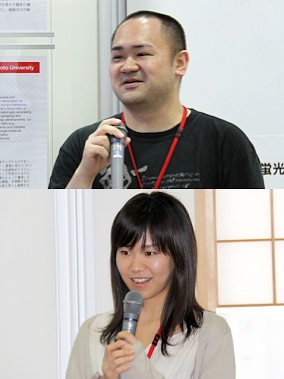
Here come the stem cells! Two young scientists, Mr. Hiroki Takeuchi (Nakatsuji Lab at the Institute for Frontier Medical Sciences, Kyoto University) and Ms. Aya Ogura (Takahashi Lab, also of the Institute for Frontier Medical Sciences) led the students through the day's investigations.
Mr. Takeuchi introduced a day in his lifestyle as a researcher in cell biology. He got a laugh, comparing his (quite clean) work area with his colleague's (rather messy) desk. Following this, Ms. Ogura explained the differences between how ES and iPS cells are created.
The mission of the day was then announced: "Are ES cells the same as iPS cells generated from skin tissue?".
To begin with, each team discussed how to investigate common characteristics of ES and iPS cells, and then used these ideas to formulate hypotheses about the day's mission.
Next, the students compared the colors, sizes, and shapes of five types of cells: mouse fibroblasts, mouse ES cells, mouse iPS cells, human fibroblasts, and human iPS cells. They found that ES cells were very similar to iPS cells in terms of their physical appearance.
Third, attention was turned to the workings of the "Nanog" gene in the ES cells. The mouse ES and iPS cells had already been specially prepared so that they would glow green when the Nanog gene was active. Observing the cells with fluorescent microscopes, the students discovered that both the ES and the iPS cells glowed green, based on which they were able to conclude that the Nanog gene was active in both types of cells.
As a final step the teams examined the cells' ability to differentiate, or change into specific cell types. They observed prepared nerve cells differentiated from human iPS cells and found that these had in fact taken on some features characteristic of nerve cells. The students also observed nerve cells that glowed green to indicate the activation of marker gene "TuJ1". The beauty of the green cell networks was admired by all.
In the end, the students realized how difficult it is to distinguish ES and iPS cells from the viewpoints of physical appearance, gene expression, and differentiation ability.
Stem cell researchers around the world are tackling this same research issue that the students spent a entire day to solve: "Are ES cells the same as iPS cells generated from skin tissue?". For the participants, the experience of being able to work on a problem at the forefront of scientific research was immensely valuable.
At the conclusion of the workshop, the students each received course completion certificates signed by Dr. Shinya Yamanaka, iCeMS professor and director of the CiRA, and iCeMS director Dr. Norio Nakatsuji.
Final Remarks
Participants' comments after the workshop indicated how impressed they were: "This is the first time I have ever had an opportunity to search on my own for an answer to a question." And: "I was moved by the beauty of the luminous cells."
The students observed that ES cells are very similar to iPS cells in many respects. Through this program, the participants learned how to make a hypothesis, test it, and then explain and organize the results.
Thank you to all who took part!
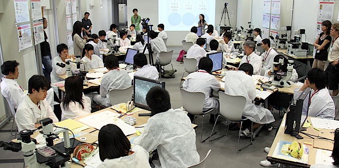 Overview of the workshop venue |
 Study Book (day 1) (PDF: 2.9MB) |
 Work Book (day 2) (PDF: 3.7MB) |
The SCG makes the above Study Book and Work Book available with the hope that numerous educators and science communicators may incorporate them into their curricula. In so doing, you are asked to respect the following:
- These texts are freely available for limited personal or not-for-profit use.
- If you wish to print a large number of copies for free distribution, or if the material in these texts will be used in a form of commercial media, then please conact the iCeMS Science Communication Group (SCG) for advance permission.
- Requests for use of individual illustrations requires the permission of the artist. Please contact the SCG for details.
email: classroom1@icems.kyoto-u.ac.jp | phone: 075-753-9784
Television Coverage
- KBS Kyoto News "KyoPlus" (August 2, 2010 17:30-)
Reporting on the teacher training that took place August 2 and 3
- Kyoto Shimbun (August 3, 2010 page 25)
- Chunichi Shimbun (August 5, 2010 evening edition page 3)
 Kyoto Shimbun (July 6, 2010 page 27)|Yahoo! News (July 5, 2010)
- Sankei Shimbun (July 6, 2010 page 23)
- Mainichi Shimbun (July 4, 2010 page 24)
- Yomiuri Shimbun (July 4, 2010 page 32)