State-of-the-art integrated imaging system allows scientists to map brain cells responsible for memory

November 18, 2014
Scientists from Kyoto University's Institute for Integrated Cell-Material Sciences (iCeMS) in Japan have developed an advanced imaging system to identify cells responsible for storing memory within a tiny worm. Their study, published in the journal Proceedings of the National Academy of Sciences, not only offers a new way to identify molecular substrates of memory but may also one day lead to understanding how memory loss occurs in humans.
The human brain is estimated to contain approximately 100 billion cells that form trillions of connections and make up a highly complex network involved in memory. To simplify the investigation of the cells directly responsible for storing memory itself, the researchers turned to a highly defined and simple worm model, called Caenorhabditis elegans, which measures one millimeter in length and has only 302 neurons.
"Worms respond to stress from vibrations by moving backwards and escaping in a process called 'mechanosensing,'" explained Takuma Sugi, who led the study, "however, over time they get desensitized to this stimulus through memory acquisition, or learned habituation."
Because only a few types of brain cells, known as neurons, in the worms are responsible for this process, it is possible to determine each cell's responsibility. But to measure the worms' movements rapidly and accurately, the scientists built a state-of-the-art integrated imaging system -- involving four charge coupled device cameras, a laser, mirrors, and an automated mechanical stimulation device -- to simultaneously capture the movement of 9 Petri dishes containing the genetically modified worms that emitted a green light upon laser stimulation.
"In the end, we identified two neurons, AVA and AVD interneurons that relay signals, to be responsible for the learned habituation," said Sugi.
Since the genome of worms is 40% identical to a human's, the scientists are hoping that the new imaging system and the interneurons they discovered may further our understanding of how memory is stored in humans.
"It is possible that these results can be applied for therapies involving memory disorders and reducing stress in the future," said Sugi.

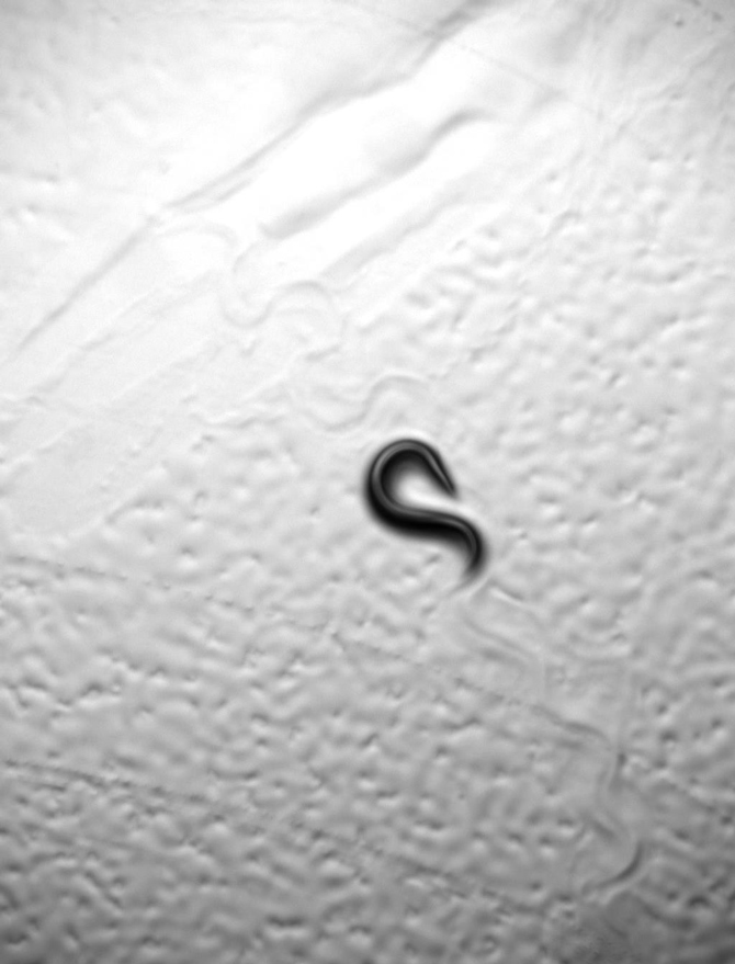
Pictured is a worm, called C. elegans, changing direction in a beautiful movement known as an 'omega turn.'
Publication Information
![]() High-throughput optical quantification of mechanosensory habituation reveals neurons encoding memory in Caenorhabditis elegans
High-throughput optical quantification of mechanosensory habituation reveals neurons encoding memory in Caenorhabditis elegans
Takuma Sugi1,2, Yasuko Ohtani2, Yuta Kumiya3, Ryuji Igarashi3, Masahiro Shirakawa2, 3
Proceedings of the National Academy of Sciences of the United States of America (PNAS) | Published Online 17 November 2014 (3PM EST)| DOI: 10.1073/pnas.1414867111
- PRESTO, Japanese Science and Technology Agency, 4-1-8 Honcho, Kawaguchi, Saitama 332-0012, Japan
- Institute for Integrated Cell-Material Sciences (WPI-iCeMS), Kyoto University, Yoshida-Honmachi, Sakyo-ku, Kyoto 606-8501, Japan
- Department of Molecular Engineering, Graduate School of Engineering, Kyoto University, Nishikyo-ku, Kyoto 615-8510, Japan
Media Coverage
- BioPortfolio
 State-of-the-art integrated imaging system allows mapping of brain cells responsible for memory (November 17, 2014)
 - Medical Xpress
 State-of-the-art integrated imaging system allows mapping of brain cells responsible for memory (November 17, 2014)
 - Science Daily
 State-of-the-art integrated imaging system allows mapping of brain cells responsible for memory (November 17, 2014)
 - e Science News
 State-of-the-art integrated imaging system allows mapping of brain cells responsible for memory (November 18, 2014)
 - Chicago Chronicle
 State-of-the-art integrated imaging system allows mapping of brain cells responsible for memory (November 18, 2014)
 - Medical News Today
 Brain cells responsible for memory mapped by state-of-the-art integrated imaging system (November 19, 2014)
