Linking Cell Biology and Materials Science through Light
Light is an indispensable tool for science. Technologies that take advantage of the different wavelengths of light can be used to study many different scientific fields.
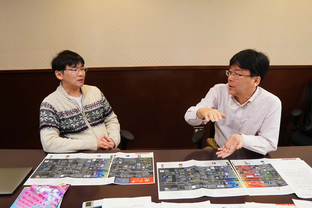
Much research at iCeMS depends on light. Here we speak with two of our scientists who are using light to observe cell biology and materials science.
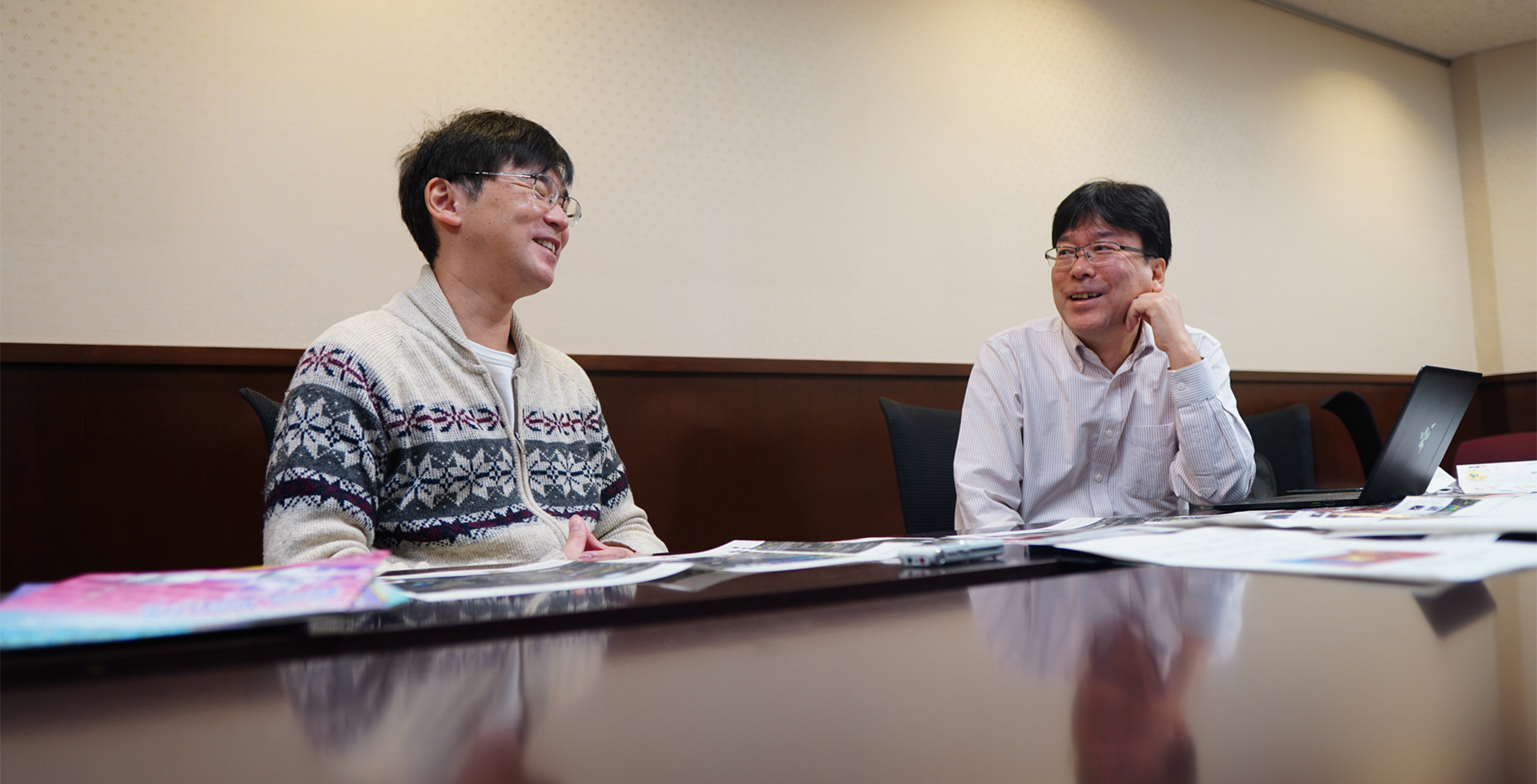
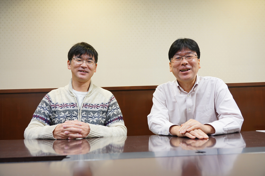
Kunihisa Sugimoto
Program-Specific Associate Professor
Born in 1972. Graduated with a PhD (Science) from the Graduate School of Chemistry, Kindai University. He was a Visiting Associate Professor at Kyoto University before taking his current position in 2019.
Takahiro Fujiwara
Program-Specific Associate Professor
Born in 1968. Completed the program of the Graduate School of Arts and Sciences, the University of Tokyo, and obtained a degree of Doctor of Science (thesis) from Nagoya University. After working as a Specially Appointed Assistant Professor at the Institute of Frontier Life and Medical Sciences, Kyoto University, he joined iCeMS in 2008 as an Assistant Professor and became a Program-Specific Associate Professor in 2017.
Public Engagement Unit (hereinafter known as “PE”)hen we talk about light, the wavelengths and technologies that are used differ according to the research field. Tell us about what type of light you are using and what kind of research you are conducting with light.
Developing technologies to see single molecules using visible light; making it possible to visualize phenomena that has been invisible up to now.
Fujiwara
In my research, I use visible light as a light source for optical microscopy. Throughout the history of biology, the optical microscope has led to many important discoveries. Through a combination of lenses, people realized that objects can be magnified. At the beginning, the light source used was sunlight, which includes visible light wavelengths. The theory and the development of technologies using light started first and foremost with visible light.
The good thing about the optical microscope using visible light is that it is a very convenient tool to do biological research. When I majored in biology in graduate school, I found it fascinating that by magnifying cells through a microscope, I could see various things moving inside the cells. I was impressed by the fact that we can observe so much activity inside cells.
PE Could you give more details about the phenomena seen with optical microscopy?
Fujiwara
Yes. My specialty is the imaging of single molecules. I observe the movements of single molecules that cannot be seen using ordinary methods. Some ingenuity is required to study how single molecules work in cells.
Without any modification, single biomolecules are usually transparent to light. So, I first used colloidal gold particles to label them. Using small particles that are a few tens of nanometers in size, the movement of single molecules can be visualized on the cell surface by utilizing the property that metals scatter light without passing it.
After around the year 2000, with the advent of GFP (green fluorescent protein) and some chemically synthesized fluorescent dyes with good photophysical properties, visualizing a single biomolecule with a fluorescent probe that is about the same size or even smaller became more practical.
In addition, technologies on visible laser light sources with high-power and stable characteristics, objective lenses with larger numerical apertures that can collect more light, and high-sensitive cameras that can detect weak levels of light have also progressed. While improving gold-particle labeling and tracking technology, the gold probe technology was combined with technologies for detecting single fluorescent probes, and I shifted my research to developing methods for single molecule fluorescence imaging in living cells.
PE Thanks to the development of fluorescent probes and the development of microscope technology, you can see more and more inside a cell. What is indispensable technology for single-molecule imaging?
Fujiwara
A technique called "Total Internal Reflection Fluorescence Microscopy (TIRFM)" has been particularly effective for observing single fluorescent molecules in cells. The technique was also put into practical use around the year 2000. The cells to be observed are cultured on a cover glass, but only up to about 100 nanometers from the cover glass can be illuminated. Since my interest is in the function of cellular plasma membranes, this technique, which allows us to see only fluorescent molecules in or close to the plasma membrane, is very useful. Taking advantage of TIRFM, I have been working on single fluorescent molecule imaging technologies to reveal the structure and function of plasma membranes.
I would like to further develop technologies to visualize phenomena yet to be seen with current technologies, and investigate the principles of the phenomena such as the movements of plasma membrane molecules, their signal transduction, and the formation of mesoscale functional structures.
PE It sounds like the technologies you mentioned come from different scientific areas, but you work on all of them?
Fujiwara
My primary job is to improve the performance of the system as a whole by integrating the properties of individual technologies to visualize single molecules. In terms of the development of a specific technology, I am collaborating with a camera manufacturer to develop high-sensitive cameras.
Technically, we are particularly focusing on high-speed observations. There are many events or phenomena that can only be seen at high speeds. For example, high-speed observations revealed for the first time that there exists a meshwork of actin filaments, the so called “membrane skeleton”, on the inner surface of the plasma membrane, and compartmentalization of the movements of membrane molecules within the meshwork regulates their functions in various ways.
Analyzing the structure of atoms using X-rays from synchrotron radiation facilities
PE Dr. Fujiwara uses visible light. But you, Dr. Sugimoto, use X-ray, which is invisible.
Sugimoto
Yes, I primarily research methodologies for crystal structure analysis using X-rays. The advantage of X-rays is that the wavelength is similar to the distance of atomic bonds in a molecule so that they interfere easily with each other.
The most interesting point in my research is that the three-dimensional structure of atoms in a molecule can be constructed by analyzing the intensity data of the two-dimensional image. A physical phenomenon called ‘diffraction’ is useful for this analysis. You probably learned in junior or senior high school about "the diffraction of light". In this class you learned that as light passes through a grating, interference occurs, and diffraction fringes are observed on a screen. Atoms act as the diffraction gratings in dependence on a crystal structure. The atomic distance and the X-ray wavelength are roughly the same. Therefore, when X-rays pass between atoms, some strengthen and others weaken, and a picture appears as a diffraction image. We are using this information for crystal structure analysis. Furthermore, the strength of the diffraction image is observed, because the atoms that form the diffraction grating have different abilities to scatter X-rays. Atoms with a higher atomic number (atoms with many electrons) diffract X-rays more strongly. Since the ability to scatter depends on the number of electrons, light elements such as carbon in the crystal are weak, and heavy elements such as metals have strong diffraction intensities. In fact, various atoms and molecules are arranged in three dimensions, and the diffraction image comes out by their combined effect.
PE X-rays are not like visible light, where you can use the sunlight, so do you need extraordinary experimental facilities?
Sugimoto I am conducting an experiment at the largest synchrotron radiation facility in Japan, SPring-8, in Hyogo prefecture. The reason why I use SPring-8 instead of the iCeMS lab is that the research I am working on is to visualize the behavior of electrons in matter. This study requires very strong and stable X-rays. It is difficult in a laboratory to accurately visualize the electron density distribution such as valence electron, which contributes only a small percentage of the total amount of electron density distribution. In order to collect data with less error, it is more advantageous to use X-rays with shorter wavelengths than in the lab. These experiments can only be done at SPring-8.
PE Short wavelengths? How short are they?
Sugimoto n my experiments, I use X-rays with a wavelength of about 0.3 to 0.4 Å (1 Å is 10 nm). One of the reasons I use short wavelength X-rays is that they have high penetrating power. For example, CT images used in sliced images of the human body are obtained by the contrast created by the difference in the X-ray absorption, which differs depending on the substance. On the other hand, in X-ray structure analysis, if there is contrast (i.e., X-ray absorption) due to the sample, an error will occur in the intensity. Short wavelength X-rays reduce that contrast. One might think that observations are easier if there is contrast, but when looking at materials in our X-ray structural analysis research, it is better to have no X-ray absorption by the sample for more accurate analysis. Of course, doctors who use CT images need the images to show contrast for diagnostics. Even if you use the same X-rays, how to use X-rays differs depending on what you want to see. In recent research, I also focus on observing dynamic structural changes like Dr. Fujiwara.
PE Instead of seeing one molecule like Dr. Fujiwara, you will be seeing at the level of one atom.
Sugimoto It is difficult to see one atom moving, but we measure the movement while the substance is playing to analyze the structure of the substance. As Dr. Fujiwara said, you can see what kind of mechanism the substance plays. In fact, the origin of the function of a substance is not so simple mechanism, and the origin often happens in a complicated manner through several steps. I think that structural analysis using X-rays has the advantage of being able to directly observe structural changes.
PE When you say “the movement”, do you mean the crystals move?
Sugimoto I guess they move. One study by Prof. Susumu Kitagawa is on the adsorption and desorption of gas on a porous metal complex (PCP). As the gas enters the pores, the crystals still move. The structure of PCP, which is said to be a gate open type, changes before and after the gas molecules are occluded. If you take snapshots of the gas occlusion process at short intervals, you may find that you cannot go to the next step unless you pass through a structure that is different from the one before and after the gas occlusion. I think that such a mechanism cannot be easily clarified without using synchrotron radiation X-rays.
PE Dr. Fujiwara, you also mentioned high-speed observations.
Fujiwara What is the speed of your data acquisition? Do they differ according to the phenomenon?
Sugimoto The X-ray free electron laser (XFEL) can measure at femtosecond time resolution and SPring-8 at picosecond time resolution.
PE Talking about SPring-8, Professor Fuyuhiko Tamanoi is also using it.
Sugimoto
There are silica nanoparticles developed by Professor Tamanoi's laboratory, and when they are irradiated with X-rays of a fairly short wavelength of 50.25 keV, electrons are repelled from the nanoparticles and can kill cancer. For some reason, this effect does not happen unless at 50.25 KeV. Auger electrons are generated even when irradiated with higher X-ray energy, but they do not act on cancer.
In principle, even at 50.3 keV, electrons are ejected in the same way, so it should work, but the electrons do not attack cancer. For some reason, they act on cancer only when irradiated with X-ray energy of 50.25 KeV. The X-rays do not attack the cancer, the Auger electrons generated by the X-rays do. At X-rays higher than 50.25 KeV, electrons will still be ejected out, but the cancer cells will not die.
I’m not sure the reason why, but Professor Tamanoi told me to think about it (laughs).
The point is to “visualize/see the things we want to see”
PE
When I hear about the research that you have explained, I think that Dr. Fujiwara's research is to see one molecule with light, and Professor Sugimoto's research is to see materials.
Even within iCeMS, there are many types of research that deal with light.
PE It seems that through research using light, both of you have placed the focus on “visualizing” things.
Sugimoto Yes, I think our common aim is to “visualize/see the things we want to see”
Fujiwara
he point is to use a wavelength that suits each purpose. We use the light required to see bigger things such as molecules and cells. You cannot use X-rays to see big things. So, when we want to investigate an unknown structure or function, we will consider "what light we should use and how it should be modulated to see what we want to see".
Dr. Sugimoto, you don’t use optical microscopy in visible light wavelengths very often?
Sugimoto I don't use it for measurements, but I use a normal stereo microscope every day. I use it when preparing samples.
Fujiwara So for you it’s a tool to prepare samples (laughs).
PE Both materials science and cell biology use stereo microscopes to prepare samples. What collaborative research are you doing at iCeMS?
Fujiwara The iCeMS Analysis Center that I am involved in running has confocal microscopes and super-resolution microscopes, and recently the group of Junior Associate Professor Ganesh Pandian Namasivayam utilized them for their research on developing probes to visualize telomere dynamics.
The fun of “seeing” with light
PE What is the appeal of using light for research and observations?
Sugimoto
When I was a student, I was originally studying the synthesis of metal complexes. Metal complexes are also used by iCeMS researchers, but you cannot write a research paper about these complexes without knowing their structures, so you first need to identify the structure. When I was in college, I enjoyed synthesizing metal complexes, but I find it more interesting to analyze their structure, so after graduating from university, I got a job as a researcher at an X-ray equipment company.
The most enjoyable thing about "seeing" with light is that we can see a world that no one has ever seen by using X-rays. Not only the metal complexes that I have made by myself, but also the crystal structures of other materials can be seen too. However, even if you look at the distribution of electrons at a glance, you can't immediately understand the structure, so it's like solving a puzzle. Solve the puzzle first and then you can see it for the first time. That's why I can see things before Professor Susumu Kitagawa can (laughs).
Fujiwara When you observe the metal complex, is it important to use imagination to interpret the data?
Sugimoto
Yes. The atoms in the crystal are symmetrically arranged. In the actual analysis, the structure is not analyzed from one end to the other end. Since we understand only a part of the smallest unit and it has some symmetry, we derive one structure by connecting it with periodic "spirals" and "folds".
But sometimes, we have to use our imagination to do it.
PE It’s like asking, “what’s going on in here?”
Sugimoto Yes. Through the symmetry, we can predict things like this could be a hexagon or something like that.
PE You will need to have a good sense of three-dimensional space.
Sugimoto Also, if you are not familiar with chemistry to some extent, you cannot analyze the structure. In the crystal structure analysis, one cannot directly see that "this part of the molecule is carbon, and that is nitrogen". We deduce the elements by looking at the distribution of the electron density, which is like a cloud spreading around an atom.
PE There is really a need for some intuition.
Sugimoto Chemically, we know "such a reaction does not occur" or "this element and this element should not bond", thus, when an absolutely impossible structure appears, I have to reanalyze it. On the contrary, if the reasoning is correct, it may be possible to analyze it in the blink of an eye.
PE It sounds very difficult. Ultimately, there is some craftmanship behind determining the structure?
Sugimoto Yes, sometimes.
PE That’s interesting how your intuition is challenged.
Sugimoto I think the observations done by Dr. Fujiwara are the same. Intuition is an important factor.
Fujiwara
In my case, I know what I expect to see, but I also see many other unrelated signals that impede the observation. So, I only illuminate where I want to see, and only extract the signal from what I expect to see.
Furthermore, I investigate various things from their movements. Since I am observing the fluid world, the movements are basically random processes based on Brownian motion. I am looking for movements that suggest something “unusual", "immobilized", or "aggregated" in the randomness.
I think a person who has never seen the movement of single molecules cannot understand what’s going on.
PE It seems difficult to tell what to see and where to see it (laughs).
Fujiwara Sometimes, I can catch a glimpse of some non-random movement (laughs).
PE It is like an “Aha!” moment.
Fujiwara
When cells are assembled to form a structure in an organism, the biased or ordered localizations of the molecules within the cells determine the proximal-distal and apical-basal axes. However, in the molecular world, thermal motion is fundamental. You can't decide the axes unless you get the molecules together or move them in a certain direction. A cell can function only when the thermal motion of molecules is properly regulated and order is given to the randomness. For example, on the membrane, three-dimensional movements can be reduced to two-dimensional movements. Inside the cell, ATP-dependent molecular motors are used to carry things in one dimension along the cytoskeleton.
It is often difficult to investigate the mechanisms at the level of single molecules without high-speed observations. We have to extract the weak signals of single molecules at high speeds in very noisy cellular environments and examine how they are regulated. It’s a lot of fun, but it’s not straightforward. We are always aiming for "the fastest single fluorescent molecule observation in the world" in living cells, and we are always pursuing the technology for this aim.
PE Is there anything that you want to see sometime in the future?
Fujiwara I want to see the movement of membrane molecules at speeds as fast as possible. In fact, we are already close to the limit from the point of view of displacements by two-dimensional diffusion. Since each displacement by diffusion and the size of the molecule itself are getting closer and closer, it’s getting difficult to distinguish the movement from the structural fluctuation of the molecule. But I still want to push the technology to its limit.
PE It will be interesting if we can see that. What about you, Dr. Sugimoto, what do you want to see?
Sugimoto I would like to observe various phenomena in femtoseconds. With SPring-8, it's still quite difficult, but FEL is little ahead in this regard (laughs).
Fujiwara I also think our technology is a little ahead, but the signal from single molecules is not sufficient.
PE
It seems like to make these observations, you need to eliminate bottlenecks and meet all sorts of necessary conditions (laughs).
Thank you so much. I am looking forward to more discoveries that come out of your research.